

RETINAL DISEASES
Home > Service Detail
APPOINTMENTS
RETINAL DISEASES
Common Retinal Conditions
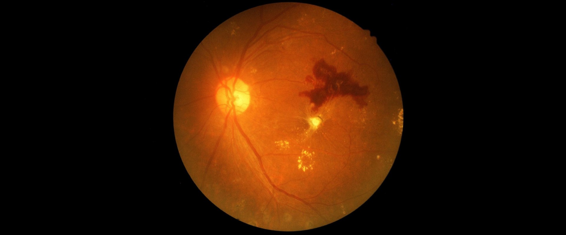
Diabetic Retinopathy
Diabetic retinopathy is a condition caused by damage to the blood vessels in the retina due to high blood sugar levels. Over time, the excess glucose in the blood can lead to the occlusion of the small vessels surrounding the retina, thus causing it to lose some of the blood supply. Consequently, the eye tries to build new blood vessels; but they are structurally abnormal and tend to leak.
To help repair the damage, an eye surgeon removes the vitreous gel from the eye to access and repair the retina, sealing any tears or leaks and replacing the gel with a saline solution or gas bubble to help the retina heal.
Early detection and management are essential to prevent vision loss.
Symptoms of Diabetic Retinopathy:
Blurred or fluctuating vision
Dark spots or floaters
Impaired color vision
Empty or dark areas in the vision
Difficulty seeing
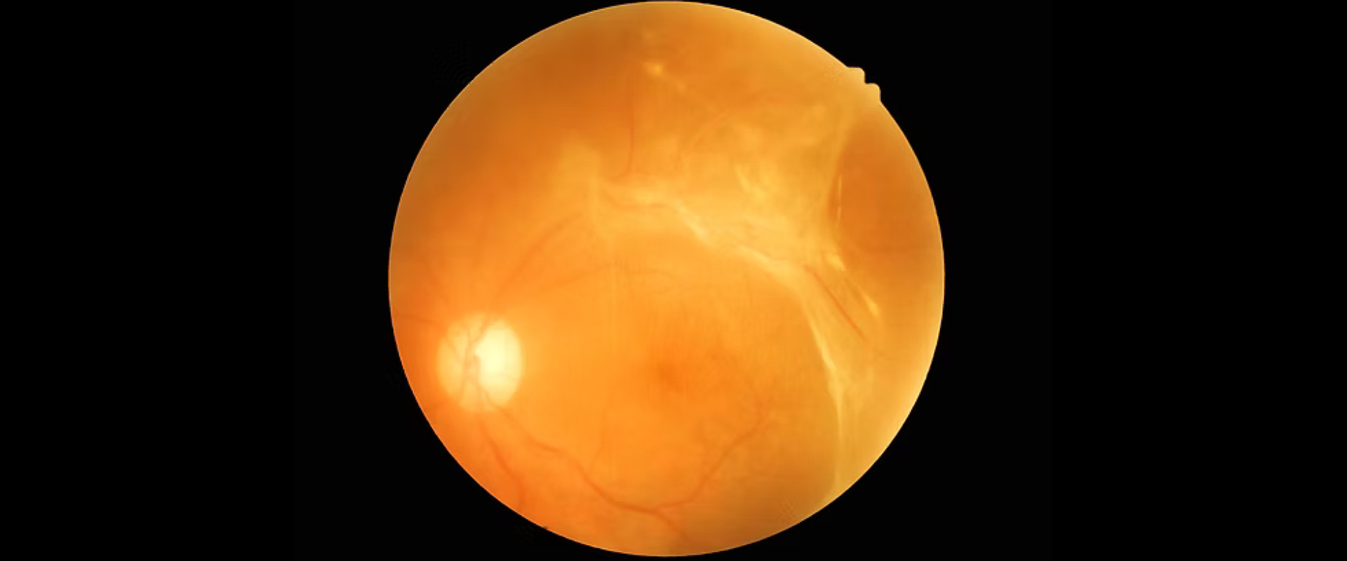
Retinal Detachment
What is Retinal Detachment?
A retinal detachment is a condition when the thin cells forming the retina pull away from the underlying layer of blood vessels, which supplies the eye with oxygen and nutrients.
In order to repair the detachment, the eye surgeon removes the vitreous gel to access the retina, repairs any tears or holes, and uses a laser or cryotherapy to reattach the retina. The vitreous gel is replaced with a gas bubble or silicone oil to hold the retina in place as it heals, restoring vision and preventing further detachment.
Some of the common causes include extreme near-sightedness, ageing, and eye injury.
Symptoms of Retinal Detachment:
Flashes of light in one or both eyes
Gradual reduction in peripheral (side) vision
The sudden appearance of floaters
A shadow or “curtain” over part of the vision field
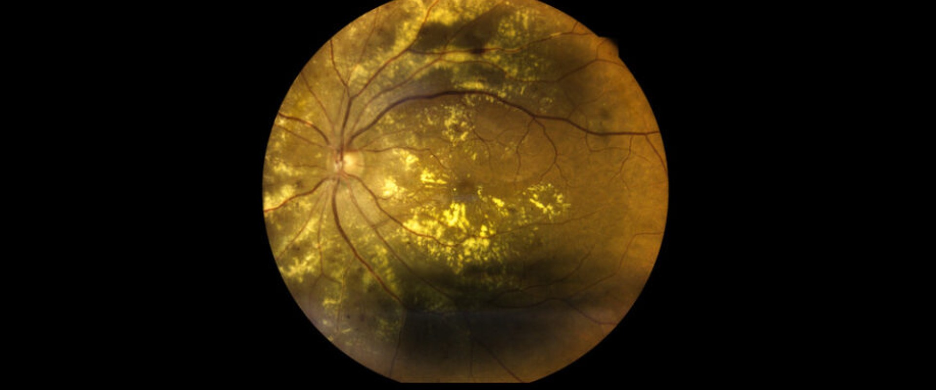
Age-Related Macular Degeneration (AMD)
AMD is a common eye condition that affects central vision, often occurring in people over 50. Some of the major risk factors include age, family history, smoking, and high blood pressure.
Treatment Options for AMD:
Anti-VEGF injections
Photodynamic therapy
Lifestyle changes and dietary supplements
Low vision aids to support vision quality
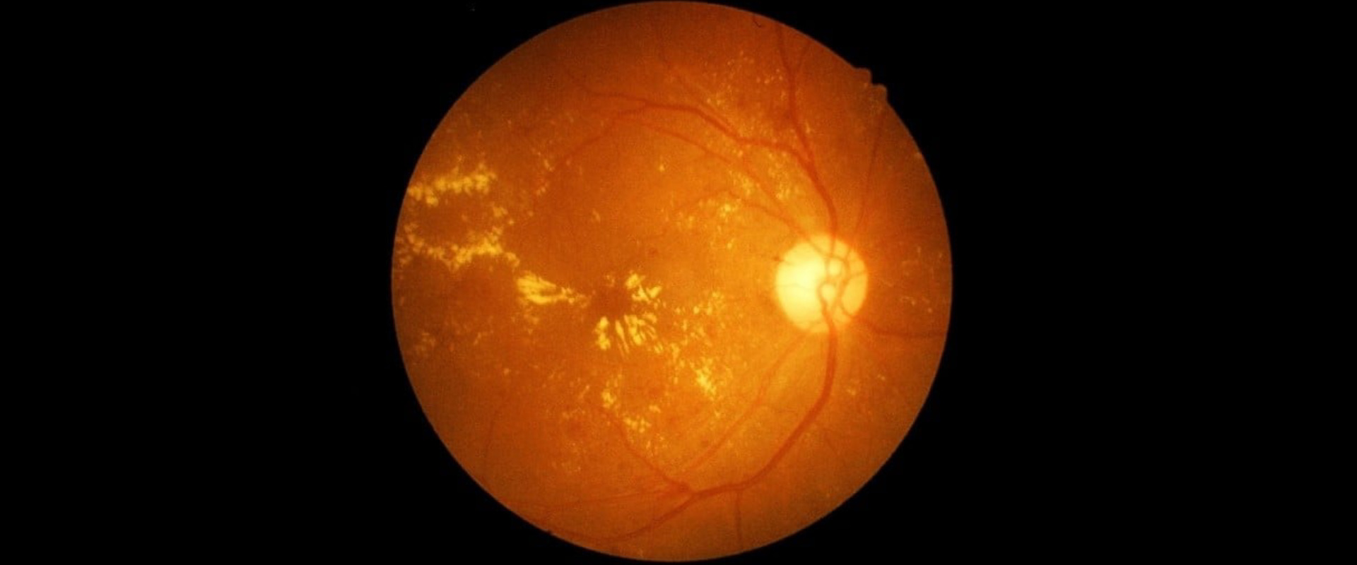
Retinal Vein Occlusion
Retinal vein occlusion is a blockage of the small veins that carry blood away from the retina, leading to swelling and vision problems. It is commonly associated with high blood pressure, diabetes, and glaucoma.
Treatment Options for Retinal Vein Occulsion:
Anti-VEGF injections to reduce swelling
Corticosteroid injections
Laser treatment to improve blood flow
Managing underlying health conditions
Advanced Diagnostic and Treatment Tools
OCT:- The imaging test that delivers high-resolution retinal layer images which help surgeons to get accurate diagnoses
Vitrectomy :- A specialized surgical procedure to remove vitreous gel, treat retinal detachment, and address other complex retinal issues.
Fluorescein Angiography:- A detailed imaging technique to assess blood flow and detect retinal blood vessel abnormalities.
Intravitreal Injections:-Medications injected into the eye to treat AMD, diabetic retinopathy, and retinal vein occlusion.
RETINA SURGERY FAQs
How many times can the retina be repaired?
Most of the time, the retina can be reattached with just one surgery. However, some people may need multiple surgeries. Over 90% of retinal detachments can be successfully repaired.
What causes the retina to weaken? Conditions like diabetes and high blood pressure can damage blood vessels and, in turn, harm the retina. Some inflammatory diseases can also cause retinal damage.
Conditions like diabetes and high blood pressure can damage blood vessels and, in turn, harm the retina. Some inflammatory diseases can also cause retinal damage.
Can the retina heal without surgery?
In rare cases, a retina can detach without a tear, often due to fluid build-up from a disease or injury. In these cases, treating the underlying condition may reattach the retina without surgery.
How is the retina checked? Eye doctors use a test called ophthalmoscopy to examine the retina, which allows them to see the retina, optic disk, and blood vessels and assess your eye health.
Eye doctors use a test called ophthalmoscopy to examine the retina, which allows them to see the retina, optic disk, and blood vessels and assess your eye health.