
 English
 العربية
 Русский
 Français
 Español
 বাংলা
বাংলা
 Kiswahili
 Deutsch
 中文
中文
 日本語
日本語
 हिंदी
हिंदी

Retinal Detachment Surgery in Delhi
Retinal detachment is a serious, sight-threatening emergency — and one that demands immediate, expert attention. When the thin layer of tissue at the back of your eye separates from its supporting tissue, every hour matters. Without timely intervention, permanent vision loss can follow.
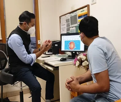
At Claritas Eye & Retina Institute, one of the most specialised Eye hospital in Delhi, patients facing retinal detachment receive world-class surgical care under the direct supervision of Dr. Mayank Bansal, a retina surgeon whose training and credentials are unmatched in the region.
Dr. Mayank Bansal Says:
“Retinal detachment is one condition where time is absolutely critical. I’ve seen patients lose sight permanently because they waited — thinking it would pass on its own. When someone walks in with flashes or a sudden curtain across their vision, we treat it as an emergency. Our goal is always to preserve as much vision as possible, and with the right technique chosen for each individual patient, we can achieve outcomes that genuinely change lives.”
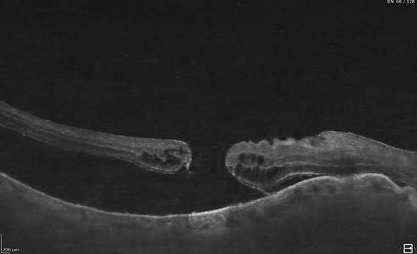
Retina detachment in Delhi is increasingly being diagnosed among all age groups from young people with high myopia to older adults with age-related vitreous changes. The good news is that with the advanced surgical techniques available today, the prognosis is excellent when treated promptly.
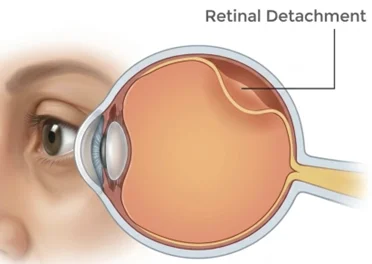
What Is Retinal Detachment?
The retina is a paper-thin layer of light-sensitive tissue that lines the back of the eye. It converts incoming light into electrical signals, which travel to the brain as visual images. Retinal detachment occurs when this layer peels away from the underlying retinal pigment epithelium (RPE), disrupting the blood supply and causing the retinal cells to begin dying.
There are three types of retinal detachment:
1. Rhegmatogenous (Most Common)
A tear or break in the retina allows fluid to seep underneath, separating it from the wall of the eye. Often linked to high myopia or age.
2. Tractional
Scar tissue on the retina’s surface pulls it away from the eye wall. Frequently seen in patients with diabetic retinopathy or previous eye surgery.
3. Exudative
Fluid accumulates under the retina without any tear or break caused by conditions like uveitis, tumours, or severe hypertension.
Sudden increase in floaters (black dots or strings)
Straight lines appearing wavy or curved
Gradual reduction in peripheral (side) vision
Flashes of light in one or both eyes
A dark curtain or shadow across part of your vision
Blurred or distorted central vision
If you notice any of these, visit a specialised eye hospital in Delhi immediately. Delay can mean permanent vision loss.
Surgical Techniques for Retina Detachment Surgery
Not every retinal detachment is the same and neither is the surgical approach. At Claritas, Dr. Bansal evaluates each case individually to determine the most appropriate technique. Three primary procedures are used for retina surgery in Delhi:
Option 01 — Vitrectomy (PPV)
The vitreous gel is removed and replaced with a gas bubble or silicone oil to push the retina back into position. Most versatile technique for complex detachments.
Option 03 — Pneumatic Retinopexy
A gas bubble is injected into the eye to push the retina back. A minimally invasive procedure suitable for selected cases with specific tear locations.
Option 02 — Scleral Buckling
A silicone band is sutured around the outside of the eye to indent the eye wall and relieve traction on the retina. Often used for younger patients with uncomplicated detachments.
Option 04 — Laser Photocoagulation
Used for early-stage retinal tears (before full detachment). Laser seals the retina to the underlying tissue, preventing progression to full detachment.
Dr. Bansal’s approach to retina detachment surgery is guided by the extent and location of the detachment, the patient’s age, lens status, and the presence of any underlying conditions like diabetes or high myopia.
Recovery & Aftercare
Recovery after retina surgery is a gradual process and differs depending on the technique used. Here is a general timeline of what to expect after your procedure at Claritas:
DAY 1 – DAY 3
Immediate Post-Operative Period
Patient will be asked to rest at home with their eyes covered. According to the procedure done, may be asked to maintain a specific head position (e.g., face-down) for few days.
WEEK 1 – WEEK 2
First Follow-Up & Gradual Return
Vision may appear blurry or foggy this is completely normal. Prescribed eye drops for anti-inflammation and infection prevention are continued. Avoid strenuous activity, bending, or lifting heavy objects.
WEEK 3 – WEEK 6
Visual Recovery Phase
Most patients notice gradual vision improvement during this period. Reading and light activities daily can usually be resumed. Regular monitoring appointments ensure healing is on track.
MONTH 2 – MONTH 3
Full Activity Resumption
Flying and certain activities must be avoided until fully resolved. Driving and normal physical activities are gradually reintroduced.
MONTH 3+
Long-Term Visual Stabilisation
Final visual acuity becomes stable. In cases where the macula was not affected, excellent vision recovery is expected. Those with macular involvement may see partial improvement over 6–12 months.
Aftercare Tips to Support Your Recovery
Use all prescribed eye drops on schedule, don’t skip doses
Avoid rubbing your eye, even if it feels irritated
Maintain the recommended head position
No air travel until your surgeon confirms
Wear UV-protective sunglasses outdoors during the healing period
Avoid heavy lifting or high-impact exercise for at least 4–6 weeks
Attend all follow-up visits even if your eye feels fine
Contact us immediately if you notice vision worsening or new floaters
Why Choose Claritas Eye Hospital for Retina Detachment Surgery in Delhi?
Choosing the right surgeon and the right eye hospital for retinal detachment is one of the most important decisions you’ll make for your vision. Here is why patients across Delhi trust Claritas Eye & Retina Institute:
World-Class Surgical Training
Dr. Bansal trained at AIIMS and UCLA’s Stein Eye Institute, bringing internationally refined surgical techniques to patients in Delhi.
Retina-Only Specialisation
Claritas is a dedicated eye and retina centre. Retinal surgery is not a side service it is the core of what we do.
Surgeon-Led, Personalised Care
Every patient is assessed and operated on by Dr. Bansal himself. No delegation to junior doctors for critical procedures.
Advanced Surgical Equipment
Our operation theatre is equipped with state-of-the-art vitrectomy systems, wide-angle viewing, and high-resolution diagnostic imaging.
International Patient Care
Claritas receives patients from across India and internationally, supported by multilingual communication and dedicated coordinators.
Research-Backed Practice
Dr. Bansal’s novel surgical technique is published by the American Academy of Ophthalmology. He actively contributes to global ophthalmology research forums.
Transparent Communication
We believe patients deserve honest, clear explanations of their condition and options, not medical jargon that leaves them confused.
Post-Surgery Support
Our care doesn’t end in the operating room. Comprehensive follow-up protocols ensure your recovery is smooth and monitored at every step.
FAQs
1. Is retinal detachment surgery painful?
2. What is the success rate of retinal detachment surgery?
3. How long does recovery take after retina surgery in Delhi?
4. Can retinal detachment be treated without surgery?
5. Who is at higher risk for retinal detachment in Delhi?
Individuals at higher risk include:
- People with high myopia (severe nearsightedness)
- Patients with a history of eye injury or trauma
- Those with diabetic eye disease
- Individuals who have undergone previous eye surgeries like cataract surgery
- People with a family history of retinal detachment
Regular eye check-ups at a trusted Eye Hospital in Delhi or a reputed Eye Hospital in India can help detect early signs and prevent complications.
Disclaimer: The information shared in this content is for educational purposes and not for promotional use.