
 English
 العربية
 Русский
 Français
 Español
 বাংলা
বাংলা
 Kiswahili
 Deutsch
 中文
中文
 日本語
日本語
 हिंदी
हिंदी

Vitrectomy Surgery in Delhi
Home > Service Detail
Vitrectomy Surgery in Delhi
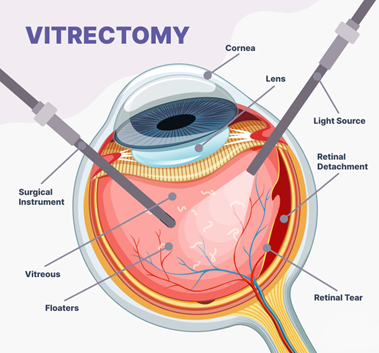
Vitrectomy is a specialized surgical procedure used to treat serious conditions affecting the vitreous — the gel-like fluid that fills the interior of the eye — and the underlying retina. During the surgery, a retina specialist removes the vitreous humor and, depending on the condition, repairs damaged retinal tissue, removes scar tissue, or treats bleeding inside the eye.
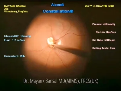
At Claritas Eye & Retina Institute, one of the leading eye hospitals in Delhi, we perform this procedure using the latest micro-incisional (23G/25G/27G) vitrectomy systems that allow for smaller incisions, faster recovery, and highly precise surgical outcomes.
Whether you are dealing with a sudden vision loss due to retinal detachment, diabetic complications causing vitreous bleeding, or a complex macular condition, timely and expert vitrectomy surgery in Delhi can make the difference between preserved vision and permanent visual impairment.
Dr. Mayank Bansal Says, “Vitrectomy is not just surgery — it is a race against time. When the retina is under threat, every hour counts. At Claritas, we have built our protocols so that patients receive a rapid, accurate diagnosis and a surgical plan the same day. Our goal is always to save and restore the maximum possible vision using the least invasive approach.”
Conditions Treated with Vitrectomy Surgery
Dr. Mayank Bansal performs vitrectomy in Delhi for a wide range of sight-threatening conditions. Each case is individually assessed using high-resolution retinal imaging before a surgical plan is created.
# | Condition | Description |
1 | Retinal Detachment | When the retina separates from the back wall of the eye, vitrectomy allows the surgeon to reattach it using gas or silicone oil tamponade. |
2 | Diabetic Vitreous Hemorrhage | Blood leaking into the vitreous due to diabetic retinopathy can block vision. Vitrectomy clears the hemorrhage and laser-treats the underlying vessels. |
3 | Macular Hole | A small break in the macula causes central vision loss. Vitrectomy with ILM peeling and gas tamponade is the standard treatment. |
4 | Epiretinal Membrane | Scar tissue on the macula distorts and blurs central vision. Surgical peeling during vitrectomy can significantly improve visual quality. |
5 | Tractional Retinal Detachment | Fibrovascular tissue pulling on the retina — common in advanced diabetic eye disease — requires vitrectomy to release traction. |
6 | Endophthalmitis | A severe intraocular infection requiring emergency vitrectomy and intravitreal antibiotics to prevent total vision loss. |
7 | Dropped Lens Nucleus | Lens material that falls into the vitreous during cataract surgery needs vitrectomy for safe removal. |
8 | Proliferative Vitreoretinopathy | Complex scarring after retinal detachment. Revision vitrectomy with membrane peeling addresses this challenging condition. |
Vitrectomy Surgery: Step-by-Step Process
Understanding the surgical journey helps reduce anxiety and improves surgical preparedness. Here is how a vitrectomy procedure is performed at our retina surgery centre in Delhi.
Step 1: Pre-operative Assessment
A comprehensive retinal evaluation including OCT (Optical Coherence Tomography), B-scan ultrasonography, and fundus imaging is conducted. Blood pressure, blood sugar, and general fitness are also checked. Dr. Bansal personally reviews all scans and discusses the surgical plan with the patient.
Step 2: Anaesthesia
Vitrectomy is most commonly performed under local anaesthesia (peribulbar or retrobulbar block) with sedation, ensuring the patient is comfortable and pain-free. In certain cases especially paediatric or highly anxious patients general anaesthesia may be used.
Step 3: Micro-Incisional Access
Three tiny sclerotomies (incisions less than 0.5 mm) are made in the white of the eye using our 25G or 27G gauge vitrectomy system. These micro-incisions eliminate the need for sutures in most cases and dramatically reduce recovery time.
Step 4: Vitreous Removal & Retinal Repair
The vitreous gel is carefully removed using a high-speed vitreous cutter. Depending on the condition, additional manoeuvres such as membrane peeling, laser photocoagulation, drainage of subretinal fluid, or placement of silicone oil or gas are performed to repair the retina.
Step 5: Closure & Recovery
The sclerotomies are self-sealing in most cases. An eye pad is applied, and the patient is moved to recovery. Most patients are discharged the same day or after an overnight stay. Postoperative instructions regarding posturing, eye drops, and follow-up are clearly explained.
Recovery & Aftercare
Typical Recovery Timeline
- Day 1-3: Mild discomfort, redness, and blurring are normal. Strict adherence to prescribed eye drops is essential.
- Week 1-2: Vision begins to slowly improve. Avoid strenuous activity, bending, and heavy lifting.
- Week 3-6: If a gas bubble was used, patients must maintain a specific head position as advised by Dr. Bansal.
- Month 1-3: Gradual visual improvement continues. Gas bubbles absorb naturally. Silicone oil, if used, may require a second surgery for removal.
- 3-6 Months: Final visual outcome is typically assessed at 3-6 months post-surgery.
Post-operative Instructions
- Use all prescribed eye drops on schedule without skipping doses
- Avoid rubbing or pressing on the operated eye
- Do not swim or submerge the head in water for at least 4 weeks
- Wear protective eye shield while sleeping as advised
- Avoid air travel if a gas bubble has been placed (gas expands at altitude)
- Attend all follow-up appointments — weekly for the first month
- Contact the clinic immediately if pain increases or vision suddenly worsens
Why Choose Claritas Eye Hospital for Vitrectomy Surgery in Delhi?
Choosing where to have your vitrectomy is one of the most important decisions you can make for your vision. Here is why patients from across India and around the world choose Claritas Eye & Retina Institute for their retina surgery in Delhi.
Sub-Specialist Retina Surgeon
Dr. Bansal is not a general ophthalmologist — he is a dedicated vitreo-retinal sub-specialist trained at AIIMS and UCLA. Vitrectomy is his core expertise, not a secondary service.
Micro-Incisional (25G/27G) Technology
We use the latest generation small-gauge vitrectomy systems for sutureless surgery, faster healing, and reduced post-operative inflammation compared to conventional 20G techniques.
Advanced Diagnostic Imaging
Our in-house OCT, wide-field retinal imaging, and B-scan ultrasonography allow for precise surgical planning and accurate monitoring of recovery — all under one roof.
Same-Day Surgical Assessment
For urgent retinal emergencies, we offer rapid same-day evaluation and, where needed, same-day surgical planning — because in retinal disease, timing is everything.
International Patient Support
We provide complete end-to-end support for patients travelling from abroad — including visa guidance, hotel recommendations, teleconsultation, and remote follow-up after surgery.
Transparent Pricing & Ethics
We believe in honest communication. Every patient receives a clear explanation of the diagnosis, surgical plan, costs, and realistic expectations — no unnecessary upselling, ever.

FAQs
1. Is vitrectomy surgery painful?
2. What is the success rate of vitrectomy surgery?
3. How long is the recovery after vitrectomy?
4. Can I fly after vitrectomy surgery?
5. Is vitrectomy performed at Claritas Eye Hospital safe for diabetic patients?
6. How do I know if I need vitrectomy surgery?
You may need vitrectomy if you have conditions affecting the retina or vitreous, such as:
- Retinal detachment
- Diabetic retinopathy
- Macular hole or macular pucker
- Vitreous hemorrhage
A detailed eye examination at a reputed Eye Hospital in India will help determine if vitrectomy is the right treatment for you.
Disclaimer: The information shared in this content is for educational purposes and not for promotional use.