 English
 العربية
 Русский
 Français
 Español
 বাংলা
বাংলা Kiswahili
 Deutsch
 中文
中文 日本語
日本語 हिंदी
हिंदी
Negative Dysphotopsia After Cataract Surgery: Causes, Patient Experience, and Solutions
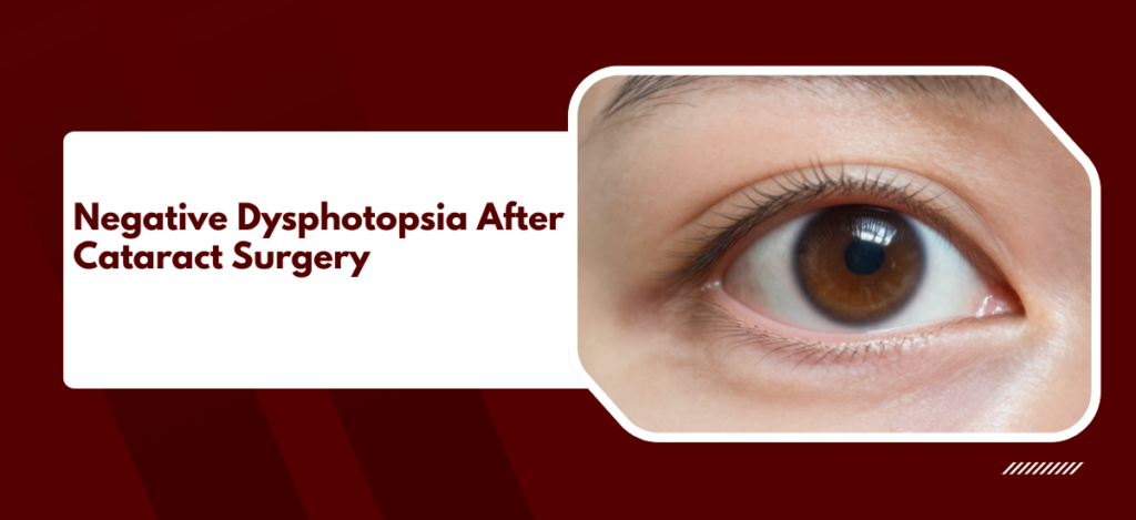
Negative dysphotopsia is a visual phenomenon reported by a subset of patients after otherwise successful cataract surgery. Patients describe a dark arc, crescent, or shadow in the temporal peripheral visual field of the operated eye, most noticeable in bright conditions. It is distinct from positive dysphotopsia, which refers to glare, halos, and starbursts caused by light scatter off IOL edges.
According to Dr. Mayank Bansal, a leading cataract surgeon at the Best Eye Hospital in Delhi,
“Negative dysphotopsia is one of the more difficult post-operative complaints to manage because the surgery has gone well by every objective measure, yet the patient experiences a genuinely bothersome dark shadow. Reassurance is the first step, but some cases do need intervention.”
Why Does Negative Dysphotopsia Happen?
The exact optical mechanism remains incompletely understood, but the most accepted explanation involves a gap in retinal illumination created by the optical geometry of modern small-diameter IOLs within the capsular bag.
- The IOL edge shadow theory: Modern foldable IOLs have a smaller diameter than the natural crystalline lens they replace. Light entering from the temporal periphery can bypass the IOL edge and fall on the anterior capsular rim in a pattern that creates a relative dark zone.
- Small pupil contribution: In bright lighting conditions where the pupil constricts, the effective optical aperture narrows and the shadow from the IOL edge becomes more pronounced rather than less.
- IOL material and refractive index: Higher refractive index IOL materials produce sharper, more defined optical edges that cast more distinct shadows compared to lower index materials.
- Nasal retinal asymmetry: The nasal retina extends further anteriorly than the temporal retina. This anatomical asymmetry means that light entering from the temporal field illuminates a zone of retina that the IOL edge can partially shadow, whereas the nasal equivalent does not produce the same percept.
- Neuroadaptation and persistence: The majority of patients experience spontaneous resolution within 6 to 12 months as the visual cortex adapts to the altered peripheral light distribution. A minority, estimated at 1 to 3 percent of cataract patients, report persistent symptomatic negative dysphotopsia beyond one year that significantly affects quality of life.
Understanding that negative dysphotopsia is an optical consequence of IOL geometry rather than a surgical error is critical for both the surgeon explaining it and the patient trying to make sense of it. For a full overview of cataract surgery and their optical characteristics at Claritas.
What Solutions Are Available for Persistent Cases?
Most cases of negative dysphotopsia resolve without intervention. When symptoms persist beyond 12 months and significantly impact daily function, a structured escalation of management options exists.
- Watchful waiting and reassurance: The first line of management for all cases is explanation and reassurance.
- Piggyback IOL: A secondary IOL implanted in the sulcus in front of the existing capsular bag IOL alters the optical geometry and repositions the shadow zone outside the functional visual field.
- IOL exchange: Replacing the original IOL with a lens of different material, edge design, or optic diameter can eliminate the shadow by altering which part of the retina the edge geometry illuminates.
- Reverse optic capture: In this technique, the optic of the existing IOL is prolapsed anterior to the anterior capsule while the haptics remain in the bag.
- Temporal YAG anterior capsulotomy: Enlarging the anterior capsulotomy at the temporal edge with YAG laser reduces the shadowing contribution of the capsular rim in the region most responsible for the percept.
Persistent negative dysphotopsia does not signify a failed surgery, but it does require a structured management pathway rather than repeated reassurance alone. For information on refractive surgery to post-cataract visual complaints.
Why Choose Claritas Eye Hospital for Negative Dysphotopsia ?
Dr. Mayank Bansal is MD (AIIMS), FRCS (Glasgow), FACS, with over 15 years of cataract and anterior segment practice including more than 12,000 cataract procedures. Pre-operative IOL selection counselling at Claritas covers optical phenomena including negative dysphotopsia, so patients understand the possibility before surgery rather than encountering it unprepared afterwards.
FAQ
Patients describe a dark arc, crescent, or shadow in the temporal periphery of the operated eye, most noticeable in bright conditions. It is not a floater and does not move with eye movement, but appears as a fixed dark zone in the outer temporal visual field.
The majority of cases resolve spontaneously through neuroadaptation within 6 to 12 months. A small proportion, estimated at 1 to 3 percent, experience persistent symptoms beyond one year that may require intervention such as a piggyback IOL or IOL exchange.
Hydrophobic acrylic IOLs with square posterior edges are more frequently associated with negative dysphotopsia than round-edge or hydrophilic designs, due to the sharper optical boundary they create at the lens margin. Higher refractive index materials also produce more defined edge shadows.
Many cases resolve without surgery through neuroadaptation. When intervention is needed, temporal YAG anterior capsulotomy is a non-incisional option with a reasonable evidence base. Piggyback IOL implantation and IOL exchange are surgical options reserved for cases that do not respond to conservative management.
*Disclaimer:* This blog is for educational and informational purposes only and should not be considered professional advice.